
پرونده:Cajal Retina.jpg
پروندهٔ اصلی (۵۰۰ × ۷۴۵ پیکسل، اندازهٔ پرونده: ۸۳ کیلوبایت، نوع MIME پرونده: image/jpeg)
این پرونده در ویکیانبار موجود است. محتویات صفحهٔ توصیف آن در زیر نمایش داده میشود. |
خلاصه
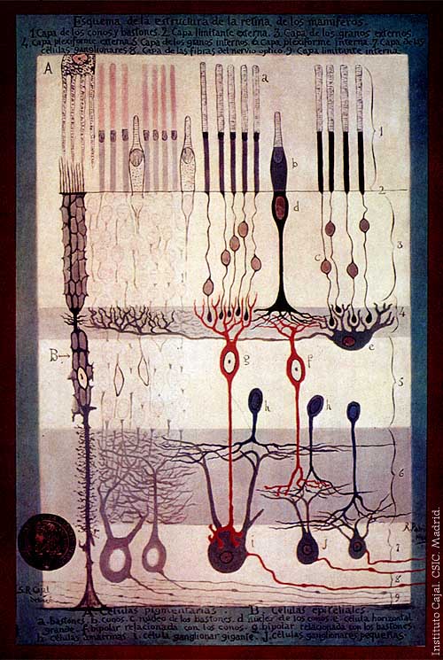
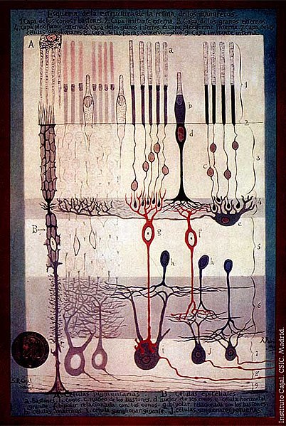
From "Structure of the Mammalian Retina" c.1900 By Santiago Ramon y Cajal.
Outline of the structure of the mammalian retina. 1. Rod and cone layer. 2. External limiting membrane. 3. Outer granular layer. 4. Outer plexiform layer. 5. Inner granular layer. 6. Inner plexiform layer. 7. Ganglion cell layer. 8. Optic nerve fibre layer. 9. Internal limiting membrane. A. Pigmented cells. B. Epithelial cells. a. Rods. b. Cones. c. Rod nucleus. d. Cone Nucleus. e. Large horizontal cell f. Cone-associated bipolar cell. g. Rod-associated bipolar cell. h. Amacrine cells. i. Giant ganglion cell. j. Small ganglion cells.
اجازهنامه
|
این اثر در کشورهایی و مناطقی که مدت زمان حق تکثیر، عمر پدیدآورنده بعلاوهٔ ۷۰ سال یا کمتر بعد از مرگ او است، در مالکیت عمومی قرار دارد. | |
| این پرونده تحت قانون حق تکثیر محدودیت آزاد منتشر شده که شامل تمامی حقوق مربوطه و حقوق نزدیک به آن میشود. | |
تاریخچهٔ پرونده
روی تاریخ/زمانها کلیک کنید تا نسخهٔ مربوط به آن هنگام را ببینید.
| تاریخ/زمان | بندانگشتی | ابعاد | کاربر | توضیح | |
|---|---|---|---|---|---|
| کنونی | ۴ مارس ۲۰۰۶، ساعت ۱۷:۴۲ | | ۵۰۰ در ۷۴۵ (۸۳ کیلوبایت) | Feezil~commonswiki | From "Structure of the Mammalian Retina" c.1900 By Santiago Ramon y Cajal. 1.- Rod and Cone layer 2.-Outer nuclear layer 3.- Granule layer 4.- External plexiform layer A: Pigmented cells; B: epithelial cells |
کاربرد پرونده
صفحهٔ زیر از این تصویر استفاده میکند:
کاربرد سراسری پرونده
ویکیهای دیگر زیر از این پرونده استفاده میکنند:
- کاربرد در ar.wikipedia.org
- کاربرد در bn.wikipedia.org
- کاربرد در ca.wikipedia.org
- کاربرد در en.wikipedia.org
- کاربرد در en.wikiversity.org
- Human vision and function/Part 1: How the eye works/1.3 Light stimulus and the eye
- User:Jtwsaddress42/People/Ramón y Cajal, Santiago
- User:Jtwsaddress42/People/R
- User:Jtwsaddress42/Gallery/Ramón y Cajal, Santiago
- User:Jtwsaddress42/Gallery/Ramón y Cajal, Santiago - The Visual System
- User:Jtwsaddress42/Gallery
- کاربرد در es.wikipedia.org
- کاربرد در et.wikipedia.org
- کاربرد در ext.wikipedia.org
- کاربرد در fr.wikipedia.org
- کاربرد در gl.wikipedia.org
- کاربرد در he.wikipedia.org
- کاربرد در hy.wikipedia.org
- کاربرد در it.wikipedia.org
- کاربرد در ja.wikipedia.org
- کاربرد در ko.wikipedia.org
- کاربرد در ml.wikipedia.org
- کاربرد در pt.wikipedia.org
- کاربرد در ru.wikipedia.org
- کاربرد در simple.wikipedia.org
- کاربرد در th.wikipedia.org
- کاربرد در uk.wikipedia.org
- کاربرد در vi.wikipedia.org
- کاربرد در zh.wikipedia.org