
پرونده:Bundleofhis.png
Bundleofhis.png (۴۰۰ × ۴۸۳ پیکسل، اندازهٔ پرونده: ۶۹ کیلوبایت، نوع MIME پرونده: image/png)
این پرونده در ویکیانبار موجود است. محتویات صفحهٔ توصیف آن در زیر نمایش داده میشود. |
خلاصه
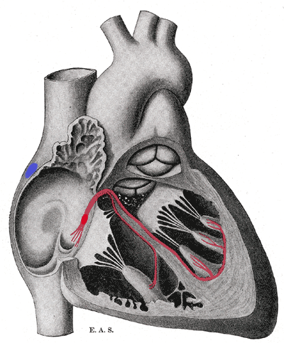
Schematic representation of the atrioventricular bundle of His. The bundle, represented in red, originates near the orifice of the coronary sinus, undergoes slight enlargement to form the AV node. The AV node tapers down into the bundle of HIS, which passes into the ventricular septum and divides into two bundle branches, the left and right bundles. Sometimes the 'left and right bundles of His' are called Purkyne or Purkinge fibres. The ultimate distribution cannot be completely shown in this diagram.
This image is misleading. Although it correctly places the SA and AV nodes in the right atrium, it appears as though there are only two papillary muscles in the right ventricle and three in the left ventricle. The opposite is actually true. The papillary muscles attach to chordae tendinae which then attach to the leaflets of the AV valves, preventing prolapse. The left AV valve is the mitral or bicuspid and only has two leaflets and therefore two papillary muscles. The right AV valve is the tricuspid and should have three papillary muscles corresponding to the three leaflets of the valve.
اجازهنامه
این پروندهٔ رسانهای در ایالات متحده در مالکیت عمومی قرار دارد. این دربارهٔ آثار ایالات متحده که حقتکثیرشان باطل شده است صدق میکند؛ اغلب به این دلیل که اولین انتشارشان قبل از ۱ ژانویهٔ ۱۹۳۰ روی داده است. برای توضیحات بیشتر این صفحه را ببینید.
|
 | |
 |
تاریخچهٔ پرونده
روی تاریخ/زمانها کلیک کنید تا نسخهٔ مربوط به آن هنگام را ببینید.
| تاریخ/زمان | بندانگشتی | ابعاد | کاربر | توضیح | |
|---|---|---|---|---|---|
| کنونی | ۲۰ سپتامبر ۲۰۰۶، ساعت ۱۷:۴۰ | | ۴۰۰ در ۴۸۳ (۶۹ کیلوبایت) | Kauczuk | Bundle of His, from Gray's Anatomy 1918 |
کاربرد پرونده
صفحهٔ زیر از این تصویر استفاده میکند:
کاربرد سراسری پرونده
ویکیهای دیگر زیر از این پرونده استفاده میکنند:
- کاربرد در ar.wikipedia.org
- کاربرد در az.wikipedia.org
- کاربرد در bn.wikibooks.org
- کاربرد در bs.wikipedia.org
- کاربرد در ca.wikipedia.org
- کاربرد در cs.wikipedia.org
- کاربرد در de.wikipedia.org
- کاربرد در de.wikibooks.org
- کاربرد در el.wikipedia.org
- کاربرد در en.wikipedia.org
- کاربرد در en.wikibooks.org
- کاربرد در es.wikipedia.org
- کاربرد در es.wikibooks.org
- کاربرد در eu.wikipedia.org
- کاربرد در fr.wikipedia.org
- کاربرد در hi.wikipedia.org
- کاربرد در it.wikipedia.org
- کاربرد در ja.wikipedia.org
- کاربرد در ja.wikibooks.org
- کاربرد در ko.wikipedia.org
- کاربرد در lv.wikipedia.org
- کاربرد در nl.wikipedia.org
- کاربرد در pl.wikipedia.org
- کاربرد در pt.wikipedia.org
- کاربرد در vi.wikipedia.org
- کاربرد در www.wikidata.org